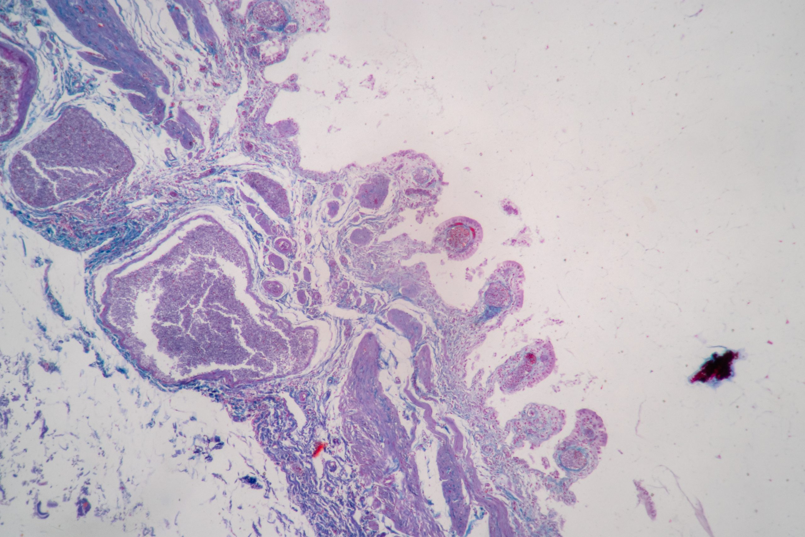
Histology is the study of tissues, their structures and how diseases impact them in distinctive ways. To study tissue, laboratories prepare and stain microscope tissue sections to show cell features for analysis. The study of how tissues are affected by diseases is called histopathology. Histopathology can help determine the severity of an illness and how far it has progressed.
C & A Scientific is passionate about making histological equipment accessible for educators and researchers. Histological analysis to diagnose diseases is a methodology that aligns with C & A’s vision to improve the health and minds of people worldwide. C & A is proud to provide equipment that enables high-quality histological research.
Histological analysis works because disease processes impact individual cells in many ways. These processes can cause them to change shape, divide, move, invade other tissues or die. These cell changes may be primary, meaning they cause the disease, or secondary, meaning the disease causes them to occur.
How are histology samples analyzed?
Histology samples must be stained. Most cells and cellular elements are almost transparent, making it tough to distinguish different cells and cellular structures if they are not stained. Typically, cells are stained with hematoxylin (or haematoxylin) or eosin. Hematoxylin stains samples blue and is alkaline, while eosin stains samples pink and is acidic.

Researchers analyzing these cells will look for proteins that signify a particular cell type or disease. These proteins are called markers. Once the tissue is sampled, it is typically placed in a solution of wax fixative to be prepared for sectioning. Histology technicians can cut the sections with various instruments, and the protocol for doing so is specific to the instrument and application. Usually, the tissue must be embedded in a medium before cutting, and in routine histology, the typical medium used is wax blocks. The wax block process requires that water be removed from the tissue while being progressively replaced by wax.
The History of Histology
Histological processes were first used in the 1600s by Marcello Malpighi, who was born near Bologna, Italy. Despite this early analysis, researchers didn’t wholly understand that organs are made of tissues until the late 1700s when Marie-Francois Xavier Bichat proposed the term “tissue” to the medical community. He suggested that one can harm tissue within organs without the organ failing. Histological staining was also first used in the late 1700s. A stain used by early botanists, called carmine, was utilized by John Hill in the 1770s to analyze tissues in ammonia solutions.
Developments in Histology
In the future, histology technicians may use artificial intelligence (AI) to enhance the histologic grading of specimens. The role of AI in histopathology may include preliminary diagnosis and quality assurance. While this advancement may see implementation soon, one downside of AI is that some diagnoses may be overlooked if a dataset is not inclusive.
If you are interested in learning more about Histology, please check out our Medical and Histology Catalogs.
C & A Scientific is a dedicated leader in improving the health and minds of people worldwide. We supply over 700 award-winning medical and STEM-inspired products to distributors and retailers looking for sensational customer service. Learn more about us and our story here.

